
I'm studying for
the NSCA-CSCS exam in June, it's comprised of two
parts. One part scientific foundations and the other part is applied. I studied
Political Science in college, not biological sciences; in fact I avoided higher
level science because math scares me. In retrospect I should have gone for it
because now I'm self studying subjects that are really nice to have a teacher
for. While studying and reviewing my materials I realized I learn best from teaching others so today we're going to talk about neurological
control of movement (how are brain tells our muscles to move).
The body has more
than 430 skeletal muscles and each skeletal muscle is an organ that contains
muscle tissue, connective tissue, nerves and blood vessels. Epimysium covers
all of our skeletal muscles and is continuous with our tendons at the ends
of the muscle. Tension is developed in a muscle because of a signals sent from
the spinal cord. Here's my understanding of how muscle contraction works.
Nerve Impluse:
Step 1:
A signal is sent
from the brain or spinal column.
Step 2: Motor
neuron in spinal column (ventral horn) is activated, and action potential
passes outward in the ventral root of the spinal cord from the nerves to
effectors (muscle)
Step 3: The action
potential in conveyed through a motor end plate on each muscle fiber
of a motor unit.
Step 4: The action
potential causes a release of acetylcholine (ACh) from the axon terminal in to
the synaptic clefts on the surface of the muscle fiber which increases the
permeability of Na+ (Sodium) in to the sarcoplasm, if there's enough ACh an
action potential will occur.
Depolarization:
Step 5: Na+ enters
muscle fiber, rapid depolarization of sarcolemma occurs= action potential
Step 6: The action
potential spreads away from the end plate in all directions and depolarizes the
Tubules where it continues down in to the sarcoplasm where is depolarizes the
sarcoplasmic reticulum (SR) membranes.
Step 7: The SR
responds to the action potential by opening Ca++ (Calcium) release channels
which floods the surrounding sarcoplasm located between the thin (actin) and
thick (myosin) filaments with Ca++.
Step 8: Ca++ binds
with Troponin, Troponin changes shape and exposes the myosin binding sites on
actin.
Step 9: Myosin
heads (cross bridges) attach to actin binding sites, the myosin head flexes
drawing actin filaments of sarcomeres towards each other. The ATP binding
site is exposed and ATP binds to the head.
Step 10: Under the
influence of ATP the myosin head detaches from actin binding site.
Steps 9 & 10
are repeated over and over again during a single contraction event as long as
ATP and Ca++ are available.
Relaxation:
Step 11: Ca++ is
returned to SR
Step 12: Troponin
again covers actin-myosin binding sites and muscle relaxes.
 More on
Muscles.....
More on
Muscles.....
Muscle cells or muscle
fibers are long and cylindrical;
they often run the entire length of the muscle and have the approximate
diameter of a human hair. These fibers have many nuclei on the edges of the
cells or fibers. Under the epimysium are in bundle groups or muscle fibers
called fasciculi,
each fasciculi is covered with a connective tissue called perimysium and each muscle fiber is surrounded by endomysium.
The endomysium is encircled by the sarcolemma or the cells membrane.
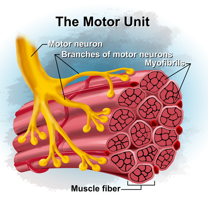
For muscle
contraction to occur there has to be a neuromuscular junction, this is
the junction between a motor neuron (nerve cell) an the muscle fiber it
innervates. One motor neuron or nerve cell can innervate several hundred muscle
fibers, but each fiber only has one neuromuscular junction. A motor neuron or
nerve cell and all of the fibers it innervates is collectively called a motor
unit.
All of the muscle
fibers of a motor unit contract when they are stimulated by the
motor neuron The extent of control a muscle has is determined by the
number of muscle fibers in a motor unit. The fewer the muscle fibers per motor
unit the more neuromuscluar control we have of those particular
movements.
The action
potential that flows along the motor neuron is not
directly responsible for creating muscle excitation, but the motor
neuron creates excitation in the muscle fiber its innervates by a
chemical transmission. When a motor unit is effected by the action potential
all of the muscle fiber with in the unit will contract. This is known as the all
or nothing principle.
There we have it,
my long winded explanation for how muscles contract, if any science people out
there want to help me out, please feel free! Thanks!
No comments:
Post a Comment